CREST Jmol Resources
Below is a collection of CREST Team Resources to help with CREST projects and materials development.
Jmol is an open source molecular visualization program available to researchers, educators and students. Although later releases of Jmol are available, they have not been completely tested for compatibility with 3D printers. We recommend using Jmol Version 14.31.36.
Jmol Version 14.31.36 (Mac or PC) (Zipped folder with Jmol.jar and Jmol.bat; use Jmol.jar)
Note that you will need to have Java on your computer to run the desktop version of Jmol. Most computers will already have a Java installed, but if you do not, you can download it for free from https://java.com/en/download/.
Digital Jmol Training Guide
These materials include and expand on the written Jmol Training materials (below) and will be replaced by the Jmol Training Guide Website (above) when it is complete.
- Getting Started in Jmol (Downloading Jmol, PDB Files, Setting Up Your Workspace, Moving a Jmol Image)
- Altering the Jmol Display (Command Line, Display Formats, Display Colors)
- Select Command and Boolean Operators
- Displaying Amino Acids (Sidechain Atoms, Bumpy Backbone, Backbone Atoms)
- Saving and Opening Files in Jmol (Saving and Opening Images; Saving, Editing and Opening Scripts, Naming Conventions)
- Adding Structural Supports (Hydrogen Bonds, Disulfide Bonds, Struts)
- Displaying Nucleic Acids (Display Formats, Coloring Schemes)
- Adding Labels to an Image (Labels, Distances Between Atoms)
- Adding Animation to Jmol (Show Orientation, Delay, Flashing, Zshade, Spin)
- Contacts (Identifying Ligand Interactions, Types of Interactions)
Written Jmol Training
A PDF version of the Jmol Training Guide that leads users through downloading Jmol, visualizing proteins using the command line and creating Jmol models to be built using 3D Printing Technology.
The written Jmol Training Guide can also be downloaded as individual sections using the links below.
Jmol Quick Reference Sheet (pdf)
A double-sided Jmol Quick Reference Sheet that highlights key commands and useful resources for designing protein models in Jmol.
How to Morph a Protein (pdf)
Instructions on how to “morph” your protein (between two PDB files) using the Yale Morph Server.
Jmol Training Videos
For those of you who want to learn Jmol by looking over someone’s shoulder, check out these videos:
- Video 1A (YouTube) – This is an 11 min video that demonstrates how to load a molecule in Jmol and how to display backbone and ligands using standard formats apropriate for building a model.
- Video 1B (YouTube) – This 17 min video demonstrates how to display sidechains (without a striped backbone), as well as using strut commands to automatically add struts within a protein. Finally, it models how to manually add struts – for ligands, ions, and other non-protein portions of your model (including DNA).
- Video 2 (YouTube) – This 21 min video addresses the fine points of adding hydrogen bonds to sheets, identifying and removing “triangle” bonds (or using code to automatically remove them), and cleaning up your design so you don’t have both hydrogen bonds AND struts in your sheets. Note that in a real model you will NEVER want purple and green for hydrogen bonds and struts! They stand out too much!
Jmol Quick Links
- Jmol Colors
- Converting Hex Colors to RGB Colors – Hex colors are accepted in Jmol by inserting an “x” inside the brackets, for example: color [xFF0000], but you may wish to use the same colors in the model description sheet, and Word uses the RGB color scheme.
Additional Jmol Notes
-
- CBM coloring schemes
- David Goodsell “soft” CPK colors (useful in distinguishing protein and substrate, or for subduing all atoms except those you want to focus on, which are presented in regular CPK)
- Carbon [211,211,211}
- Nitrogen [160, 210, 255]
- Oxygen [255, 185, 185]
- Yellow [255, 255, 100]; Salmon [255, 150, 150]; Green [0, 255, 60]
- Using PowerPoint themes or any one of a number of color palette generators may help you identify a pleasing color scheme. Coolors.co has buttons that allow you to see what a colorblind person would see.
- David Goodsell “soft” CPK colors (useful in distinguishing protein and substrate, or for subduing all atoms except those you want to focus on, which are presented in regular CPK)
- Displaying hydrogens in a pdb file
- Before loading pdb file, enter command:
- set pdbaddhydrogens true
- This will display all hydrogen atoms and will also display double bonds.
- Before loading pdb file, enter command:
- Automated removal of triangle bonds; enter command:
- for(var i=1; i<=1000; i++){var j=i+2; select resno=i or resno=j; hbond 0.0};
- Saving pdb files (and MORE) in Jmol
- Morphing two versions of a protein: Instructions are provided on this website http://www2.molmovdb.org/wiki/info/index.php/Morph_Server
- Dealing with two orientations of a sidechain
- Occasionally, a residue within a structure is found in one of two orientations. Both orientations are included in the pdb file. If you display the residue, it will display BOTH versions – which is confusing.
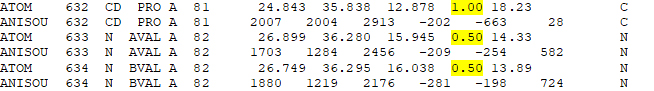
- If you download and open the pdb file in Word (adjust margins to narrow – it will be easeier to read!) and scroll to the residue, it will look like this:
- CBM coloring schemes
-
- The content highlighted in yellow indicates the percentage of time the atom is in this particular location. Typically the value is 1.00 (all the time); but if the residue is in two different orientations, the value is 0.5. If this is the case, you want to select one value to display. The primary citation will often guide you as to which orientation to select.
- Let’s assume we want to display the first orientation. In this example, the residue is val82 of chain A. The selection command to display AVAL (and not BVAL) is: Select val82 and :a and (sidechain, alpha) and %A
Guidelines for Submission
When submitting a model design for review, please include
- Model design (one of the following):
- Jmol-created jpg + copy of pdb file you used
- Jmol created png
- Script file, including the name of the pdb file OR a copy of the pdb file if it has been modified
- Model Description Sheet
- Abstract
The model should be reviewed by your instructor and/or research mentor for accuracy of displayed details; CBM staff will review the model for structural soundness.
Guidelines for submissions are provided below:
Model Design Parameters
This form will help you think through what features you might want to include in your model design.
Model Description Sheet
Each model that is built is accompanied with a Model Description Sheet that includes an abstract describing the molecular story as well as a key that describes the coloring scheme and displayed sidechains for the model. The Model Description helps to convey the molecular story of the model when you are not present to tell the story yourself.
Guidelines for Writing an Abstract
Recommended design guidelines for writing an abstract that will be included on your Model Description Sheet..
Model Design Review
Parameters to review to ensure your model is ready to build.
Guidelines for Designing a Poster
Recommended design guidelines for writing a CREST Team poster.
Jmol Images for Posters
Tricks for getting high resolution Jmol images
Sample Posters
A collection of sample posters that represent the good, the bad and the ugly.
Jmol Explorations
Many CREST Teams develop interactive molecular web pages using the molecular visualization program Jmol. Two main applications, Proteopedia and the CBM’s Jmol Exploration Creator allow for quick and easy Jmol web page development.
David Canner’s PowerPoint Presentation
Proteopedia is a free, collaborative 3D-encyclopedia of proteins & other molecules. This overview Powerpoint slideshow introduces Proteopedia and explains its basic functionality.
Jmol Exploration Creator (JEC)
In this interactive environment, users can create interactive Jmol web pages using a collection of simple buttons and controls. These tutorials can then be exported for future viewing or editing.
Viewing Small Molecules From the NIH Cactus Database: If you want to view a small molecule in the Electronic Poster Creator, enter ANY pdb file in the “Enter pdb file to be used here” box. (4ins is a small file that loads quickly.) Then, for the first command in the Jmol command box, enter:
load $filename
Some examples:
load $adenine
load $lysine
load $glucose
load $aspirin
NOTE: The CBM Jmol Explorations Creator (JEC) is hot off the press – and we are still troubleshooting. If you encounter difficulties, please contact Mark Hoelzer (hoelzer@msoe.edu). Please describe the issue as well as the browser you are using and, if possible, attach the in-progress Jmol tutorial or electronic poster being developed. We also welcome feedback on how you are using the tool!