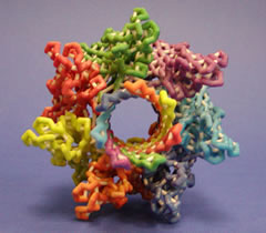
A plaster model of the protein Hemolysin.
As you explore the how the structure of the protein you are studying relates to its function, you’ll get a clearer idea of the portions of the protein you want to feature in your model. You might design a backbone model with a few key sidechains in ball and stick (spacefill and wireframe, in the parlance of Jmol). Perhaps you’ll feature sidechains important in binding with a substrate, cofactor, or another protein.
As you design your model, you should consider how the model will be used — both by the research lab and in the classroom. You may even decide that you need to design different models for the two groups. Use bright colors to draw the eye to the key features of the model; softer colors are great for more subtle detail (perhaps for distinguishing subunits). Use white (or a very light color) for hydrogen bonds and struts — these are structural features and not the main focus of the model.
No single model will adequately tell the WHOLE molecular story. Jmol explorations, a video clip (with model in hand), or an animation may be your best option to guide others through the intricacies of your protein. You may decide that two different models will be beneficial in conveying the concepts. Think creatively! Remember that CBM staff are available to discuss your ideas and provide feedback. Contact (crestprogram@gmail.com) for more information.
Go to the Jmol Resources page to download Jmol or access training materials for using Jmol.