Goucher College (Baltimore MD)
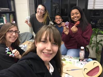
Hello! We are the Goucher College BCMB Club’s CREST team in Baltimore, Maryland. Our members consist of Dr. Judy Levine, Kelly Budge, Shannara Bauer, Brandon Creed, Olivia Dickert, and Edna Ferreira. We are all seniors here and began our school’s chapter last fall. We are very excited to join the modeling project this year. We plan on modeling OGA interacting with the inhibitor Thiamet-G which has been shown to reduce symptoms of Alzheimer’s Disease in a mouse model.
O-GlcNAcase (OGA) with Thiamet-G
PDB file: 5un9
Primary Citation: B. Li, H. Li, L. Lu, J. Jiang, Structures of human O-GlcNAcase and its complexes reveal a new substrate recognition mode. Nature Structural & Molecular Biology. 24, 362 (2017).
Abstract: Alzheimer’s Disease is associated with the hyperphosphorylation of the microtubule-associated protein tau. Protein phosphorylation events often occur at the same positions on a protein as O-GlcNac modifications. The mutually exclusive events exist in a dynamic equilibrium that is regulated by enzyme activity. O-GlcNAcase (OGA) is an enzyme which removes O-GlcNacylation from proteins. Thiamet-G was designed to cross the blood brain barrier as a selective inhibitor of OGA in order to effectively decrease the removal of O-GlcNAcylation events. By inhibiting the ability of OGA, there is evidence that Thiamet-G can lead to a decrease in hyperphosphorylation of tau in vertebrate brains and potentially serve as an effective treatment of Alzheimer’s Disease. We modeled the inhibition of OGA by Thiamet-G to highlight the enzyme residues that interact with the inhibitor and the parts of the protein that are important for recognizing target proteins.
Lane College (Jackson TN)
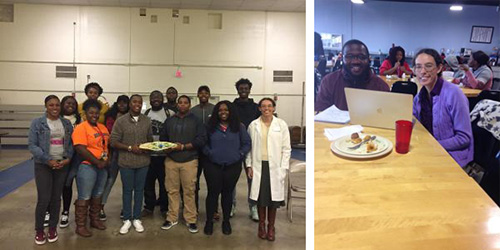
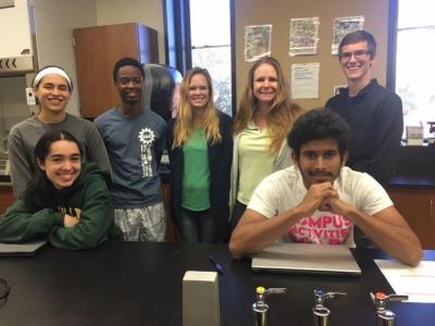
Greetings. Lane College Science Club joined the ASBMB Student Chapters last year. Our club participates in a few projects, including outreach at Lincoln Elementary School. In this photo, we just finished a program with fourth graders demonstrating Euglena and phototaxis. We are excited to be working on the CREST project building a model of human O-GlcNAcase. Our team members are Le’Juan Berry (Senior), Ashley Gayle (Senior), Darrius Hawkins (Senior), Ryan Billings (Freshman), Ashlee Martin-Richardson (Freshman), and Ihouma Tasie (Senior). Our faculty advisors are Dr. Melanie Van Stry and Dr. Candace Jones.
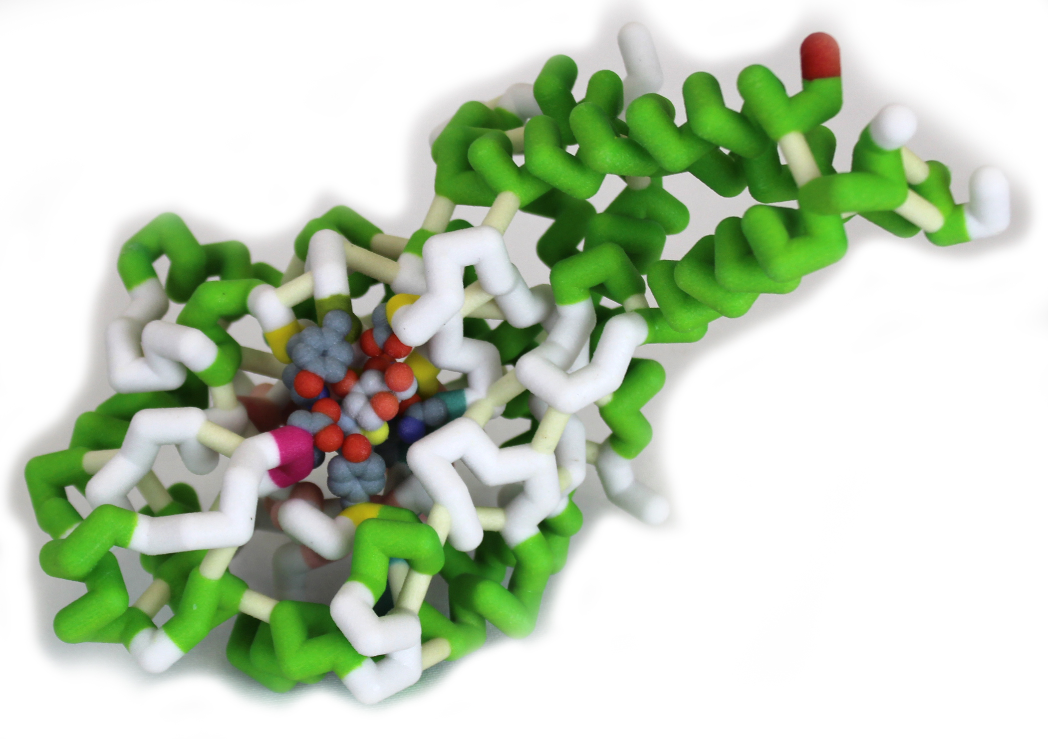
Human O-GlcNAc Hydrolase (hOGA)
PDB File: 5m7s
Primary Citation. Roth C, Chan S, Offen WA, Hemsworth GR, Willems LI, King DT, Varghese V, Britton R, Vocadlo DJ, Davies GJ. Structural and functional insight into human O-GlcNAcase. Nat Chem Biol. 2017 June; 13(6): 610–612.
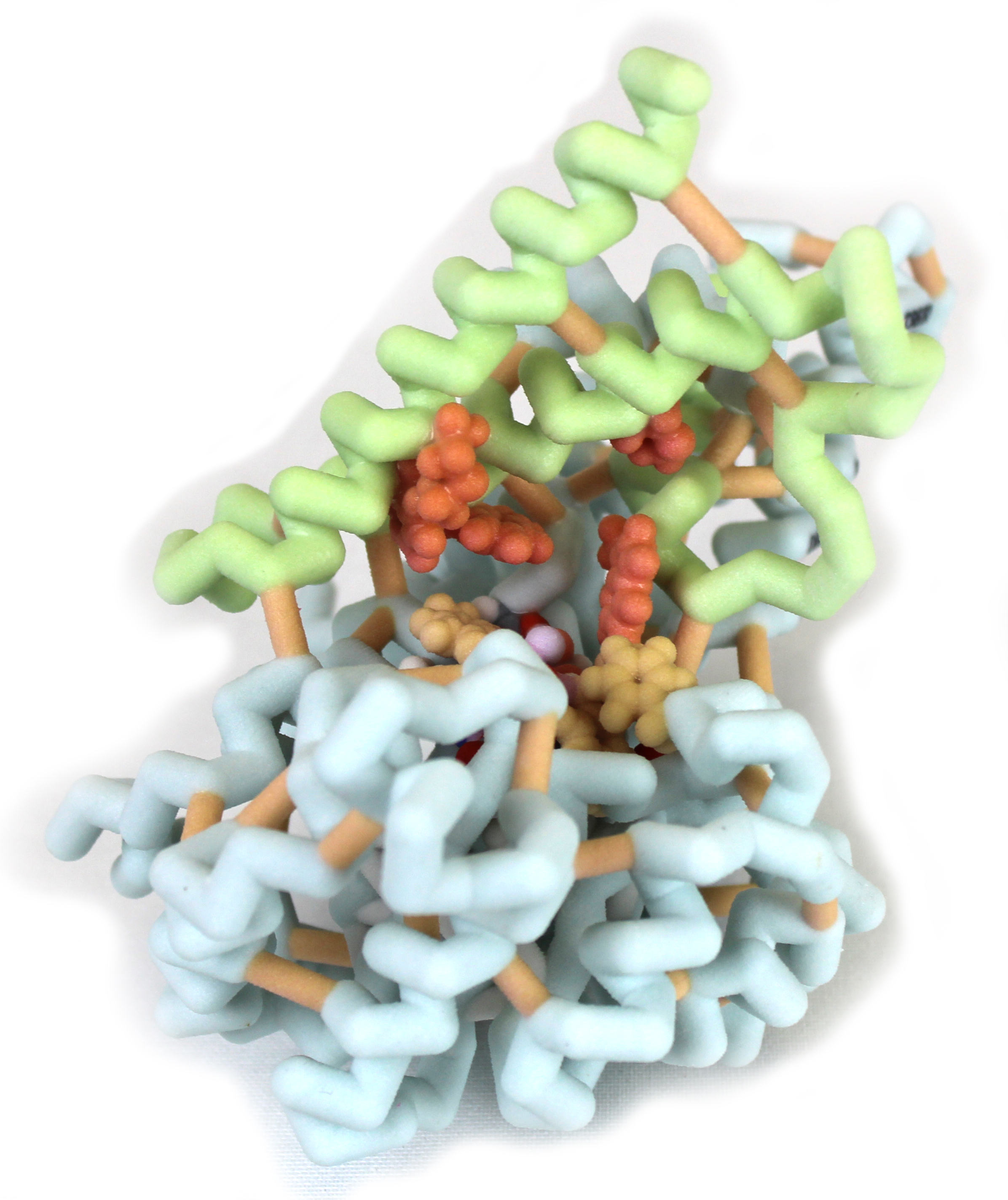
Abstract: The post-translational modification (PTM) of proteins enable cells to react promptly to internal and external signals through direct and progressive control of protein function. Addition of O-linked-n-acetylglucosamine (O-GlcNAc) to serine and threonine residues of cytoplasmic, nuclear and mitochondrial proteins is a nourishment and stress response PTM. O-GlcNAcylation has been linked to several diseases such as diabetes and cancer. For example, increased O-GlcNAcylation is directly linked to insulin resistance and to hyperglycemia-induced glucose toxicity, two characteristics of diabetes and diabetic complications. O-GlcNAc transferase (OGT) and O-GlcNAcase (OGA) are enzymes that control the dynamic cycling of this PTM. Here, we have constructed a 3D model of human OGA bound to the transition state analog Thiamet-G using Jmol and the PDB file 5M7S, a truncated version of the enzyme called Split1 (Roth et al. Nat. Chem. Biol. 2017). Split1 forms a functional homodimer that is stabilized by a helical bundle formed by helices from each subunit. Thiamet-G fits in the substrate binding pocket formed by Cys215, Tyr298, and Trp278 and interacts with Gly67, Lys98, Asn280, Glu295, and Asn313 to form hydrogen bonds. Thiamet-G also interacts with the catalytic residues Glu174 and Glu175. The focus of this project was to explore the protein structure of OGA and design and build a physical model that illustrated key functional features of the protein. Funded in part by NSF-DUE 1725940 for the CREST Project.
Nova Southeastern University (Fort Lauderdale FL)
Hello Everyone! We are a group of three students working with our faculty member, Dr. Emily Schmitt Lavin. We just became a chapter of ASBMB this fall with a small group of students. We are looking forward to working on the O-GlcNAcylation modeling project with everyone on the CREST Conversation team. Our group consists of three pre-med students: Sophia Nguyen and Vivian Perez Hernandez who are seniors, and Alesa Chabbra who is a freshman.
OGT in complex with UDP and fused substrate peptide TAB
PDB file: 5lvv
Primary Citation: Rafie K, Raimi O, Ferenbach AT, Borodkin VS, Kapuria V, van Aalten DMF. 2017 Recognition of a glycosylation substrate by the O-GlcNAc transferase TPR repeats. Open Biol. 7:170078. http://dx.doi.org/10.1098/rsob.170078

Abstract: O-GlcNAcylation is a post-translational modification similar in importance to the mechanism of phosphorylation in its ability to affect signal transduction. This process is mediated by the enzyme, O-GlcNAc transferase (OGT). OGT catalyzes the addition of the sugar, N-acetylglucosamine (GlcNAc) from the carrier molecule uridine diphosphate N-acetylglucosamine (UDP-GlcNAc) to certain serine or threonine residues in more than a thousand target substrate proteins. The TAB1 (transforming growth factor-beta-activated kinase 1 binding protein) substrate was fused to OGT. GlcNAc was shown binding to three serine residues in TAB1. Six tetratricopeptide (TPR) repeats were identified. These alpha helical paired repeats fold together to produce a single domain called the TPR domain near the N terminus of OGT. Within the TPR domain, five Asparagine (Asp) residues were identified that are involved in holding the substrate in place. Beta sheets in OGT were also indicated. If TAB1 does not receive GlcNAc it will not be able to signal the proper response of the innate immune system.
Saint Leo University (St. Leo FL)
Greetings from Saint Leo! Our team consists of our faculty member Dr. Audrey Shor and six students. Saint Leo has been a chapter of ASBMB since 2011, however this year’s student group is completely new. It includes Corey Gallen, a senior, Allison Cobb, a junior, and Anapaula Rios-Rosales, Isabella Jacus, Raheim Grant and Zarrar Rashman, all first year students. Saint Leo’s team is interested in modeling the O-GlcNAcase protein. We look forward to working on this project and with all other teams!
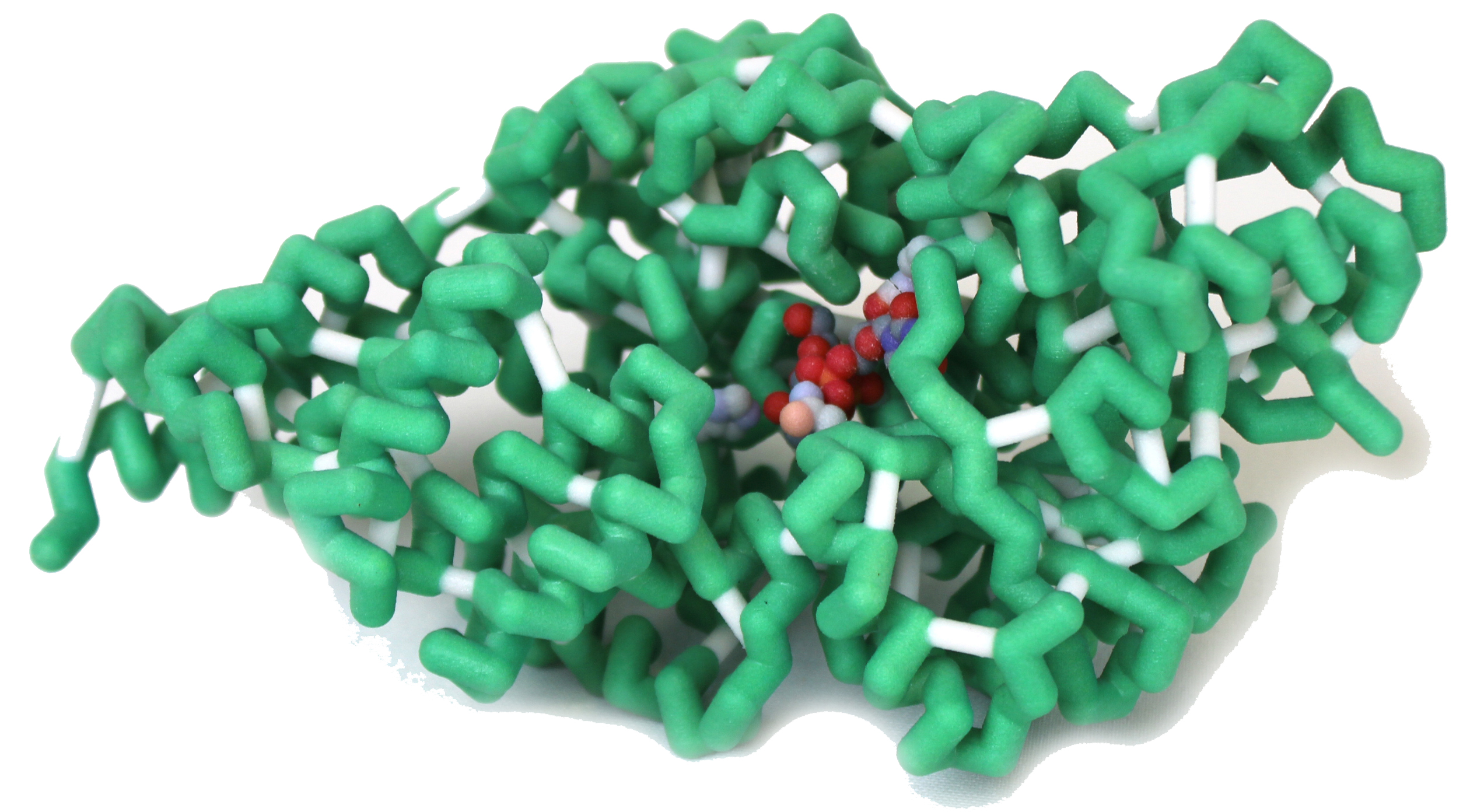
O-GlcNAc Hydrolase (OGA) with bound transition state analog ThiametG
PDB file: 5m7s
Primary Citation: Roth C, Chan S, Offen WA, et al. Structural and functional insight into human O-GlcNAcase. Nat Chem Biol. 2017;13(6):610-612. doi:10.1038/nchembio.2358.
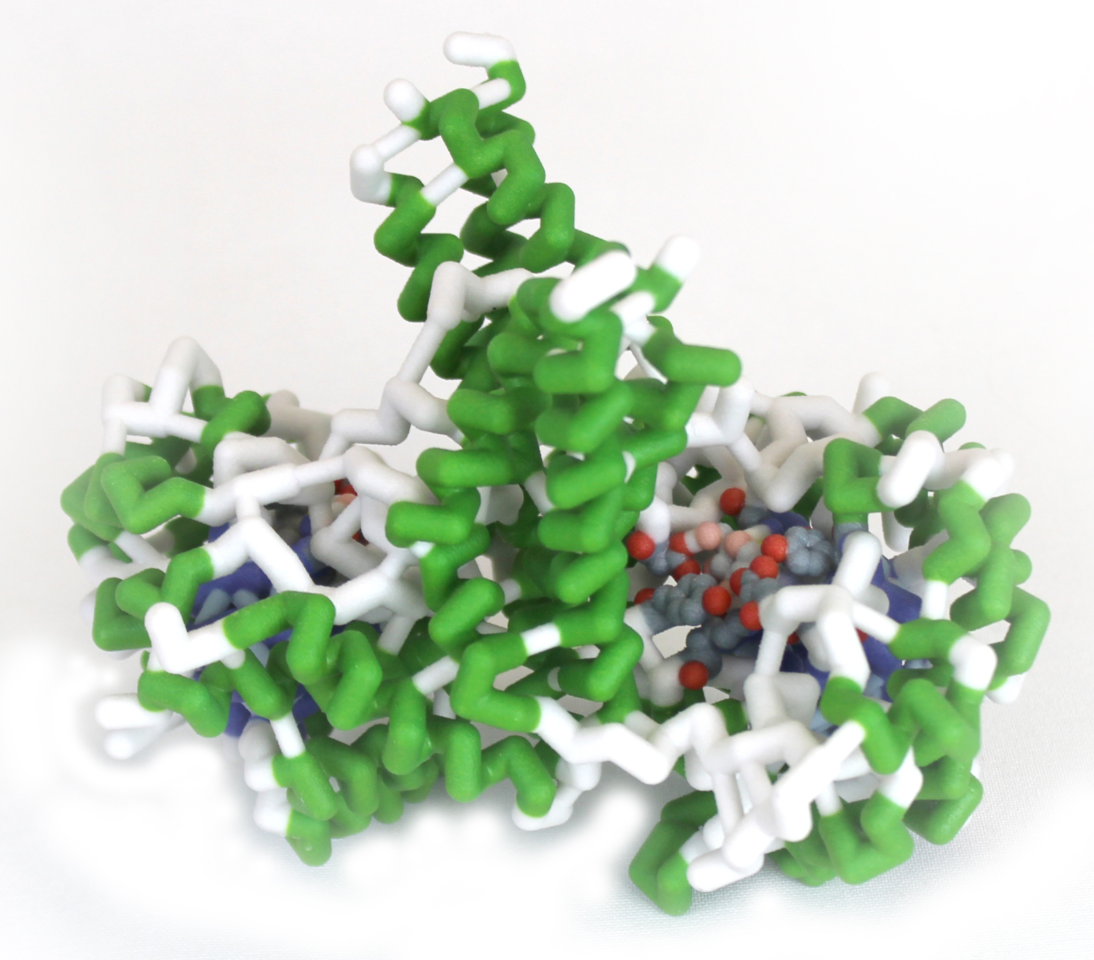
Abstract: A basic component of learning undergraduate biology concepts is grasping the significant interplay between carbohydrates and proteins. O-GlcNacylation is a post-translation modification that covalently attaches O-linked N-acetylglocosamine (O-GlcNAc) moieties to cellular proteins. O-GlcNAc is a sugar moiety that can posttransationally modify serine and threonine residues of target proteins. Much can be gained from studying the targets of O-GlcNacylation. Two enzymes regulate O-GlcNacylation; O-GlcNAc transferase (OGT), which catalyzes the addition of GlcNAc to specific serine and threonine residues, and O-GlcNacylase (OGA), which catalyzes the hydrolysis of the monosaccharide from the substrate. The protein we emphasize, OGA, is a homodimer, with each subunit comprised of 916 residues. Two isoforms of the enzyme exist in humans; 1 and 3 which are expressed in the cytoplasm and nucleus, respectively and are speculated to have different sensitivities to substrate based on their environment. The cytoplasmic isoform includes a full length protein (OGA-L), while the nuclear isoform is shorter (OGA-S), lacks a C-terminal acetyltransferase-like domain and demonstrates deduced enzymatic activity in vitro. Human OGA possesses a fugitive substrate binding groove that is highly conserved among metazoan OGAs, in addition to a stalk domain near the active site. Residues significant to O-GlcNAc, a hydrolase, are close to the sugar/catalytic machinery or occupy positions shown to undergo a conformational change upon inhibitor binding. Mutations targeting the substrate binding groove identified residues that are not required for hydrolysis but serve roles in recognition and binding of substrate. The differences between the residues required for the turnover of these substrates may reflect a difference in molecular movement in OGA sites. Further elucidation of this mechanism could help advance our treatment and/or prevention of diseases like neurological disorders and diabetes.
University of Minnesota Rochester (Rochester MN)
We have been working on our project since early in the 2017 fall semester. We are excited about our CREST project. Our team is of 8 students who have a love and curiosity in biochemistry and medicine! Here’s a picture of our tiny university in downtown Rochester since I don’t have a group picture.
PDB file: 4gz5
Primary Citation: Lazarus, M., Jiang, J., Gloster, T., Zandberg, W., Whitwort, G., Vocadio, D., & Walker, S. (2012). Structural snapshots of the reaction coordinate for O-GlcNAc transferase. Nat. Chem. Biol., 8, 966-68. Doi: 10.1038/nchembio.1109
Abstract: Currently in the United States more than five million individuals live with Alzheimer’s disease (AD), which can lead to memory loss, difficulty with problem solving, a decreased quality of life and eventually death. AD is initiated by the formation of amyloid plaques in the extracellular space of the brain, however, the reason for this is unknown. The amyloid-ꞵ precursor protein (APP), an integral membrane protein concentrated in neural synapses, has been linked to the development of these plaques. In healthy individuals, the cleavage of APP leads to a long, secreted form of APP (sAPPα) and C-terminal fragments (CTFs). In individuals with AD, cleavage of APP leads to sAPPꞵ, different CTFs, and amyloid-beta (Aꞵ) fragments. The misfolding of Aꞵ contributes to the development of AD when these Aꞵ peptides aggregate and form amyloid plaques. Previous research demonstrated that addition of O-linked N-acetylglucosamine (OGlcNAc) to Thr576 on APP decreases the number of Aꞵ peptides formed, thus O-GlcNAcylation of this protein could be useful for therapeutics and warrants further research. OGlcNAcylation occurs when the enzyme, OGlcNAc Transferase (OGT) (PDB ID: 4gz5), catalyzes covalent attachment of an N-acetylglucosamine molecule to either a serine or threonine residue via a uridine diphosphate N-acetylglucosamine (UDP-OGlcNAc). The UDP enables the transfer mechanism by acting as a good leaving group. Our model depicts OGT bound to UDP-OGlcNAc via Gln839, His498, and Lys898 that will attach OGlcNAc to Thr576 on a flexible toober that models APP. The APP toober, will demonstrate the cleavage and folding of Aꞵ peptide.