

Many Jobs of Proteins
This Jmol Exploration was created using the Jmol Exploration Webpage Creator from the MSOE Center for BioMolecular Modeling.
This tutorial focuses on the wide range of activities accomplished by proteins in cells. This section is an introduction to a variety of ways of depicting proteins, as well as some common color schemes used in depicting proteins. All the proteins used in this tutorial have had their structures determined using X-ray crystallography or NMR. These structures are deposited in the Protein Data Bank (PDB) for use by researchers, educators and students. The PDB accession numbers are provided for each protein in this tutorial, in case you want to explore the proteins further.
Throughout the tutorial, clicking on the buttons will launch a Jmol image of the protein on the screen at the right.
Once a protein appears in the Jmol window, wait until the image stops moving before pushing a button to execute another script. You may spin the protein (left mouse button click and drag), or, if you wish, you may use Jmol commands to explore the protein further. A Jmol Quick Reference Sheet and Jmol Training Guide are available if you want to play with the images. (WARNING: This can be a lot of fun!)
If you hover over an atom in Jmol, a popup window will identify the atom in the format:
[ASN]108:B CA #1888
where:
[ASN] is the 3 letter amino acid abbreviation (or 1 letter nucleotide, if it is a nucleic acid)
108: is the residue number
B identifies the chain name in the pdb file
CA identifies the atom type. (CA is the code for the alpha carbon)
#1888 identifies the atom number in the pdb file.
There are three common formats for displaying proteins; each is used to show different features of the protein. Shown here is insulin (2hiu.pdb), a hormone that regulates blood sugar levels and one that you will see again later in the tutorial.
Spacefill depicts all atoms at their relative diameter.
Spacefill PDB ID: 2hiuA backbone display only shows the central carbon atom (the alpha carbon) of each amino acid, with a solid line connecting adjacent amino acids. It is sometimes called the alpha carbon backbone.
Backbone PDB ID: 2hiuBall and stick shows each of the atoms as balls, with sticks to indicate the bonding between atoms.
Ball and Stick PDB ID: 2hiuBelow, all three depictions are displayed in succession.
Combination Display PDB ID: 2hiuRecord an advantage and a disadvantage of each type of display.
Color can be used effectively to communicate information about protein structure.
In this tutorial, the secondary structure of each protein will be color-coded. The alpha helices will be colored salmon and the beta sheets will be colored light yellow.
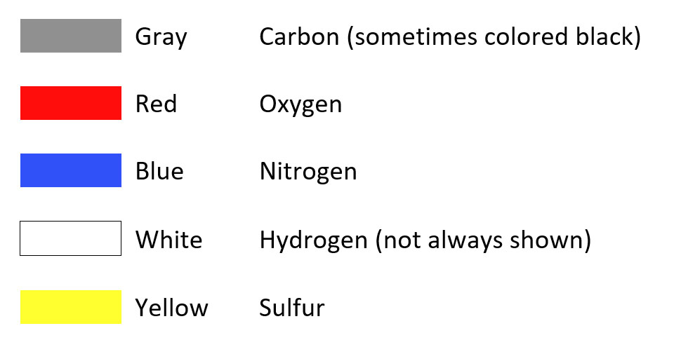
The CPK coloring scheme is a standardized coloring scheme that uses a specific color for each type of atom. In this scheme:
Click the button below to see the coloring of the secondary structure and the CPK-colored sidechains on the amylase protein (1ppi.pdb).
Sample of Color Scheme Used in this Tutorial PDB ID: 1ppiAmylase is a digestive enzyme that catalyzes the breakdown of starch into sugars. Amylase is found in human saliva, where it begins the digestive process.
Molecule of the Month for Alpha-Amylase
Color Scheme:
Protein backbone – white
Alpha helices – salmon
Beta sheets – light yellow
CPK coloring for amino acids (carbon is gray, oxygen is red, nitrogen is blue)
Light CPK coloring for sugar substrate
Calcium ion – purple
Chloride ion – lime green
Hydrogen bonds – white
In the next image, the carbohydrate substrate flashes in cyan and 'soft cpk' (light gray carbon, pink oxygen). Using light cpk allows distinction between substrate and protein.
Carbohydrate Substrate in Active Site PDB ID: 1ppiThe amylase protein contains two ions: calcium and chloride. Click the following two buttons to see the locations of each of these ions.
View of the Chloride Ion (lime green) PDB ID: 1ppiThis next section is about amylase and the properties and arrangement of its amino acids. In the first button, the hydrophobic amino acids are colored yellow (including their backbone) and the hydrophilic amino acids are colored red (including their backbone). The Amino Acid Starter Kit coloring scheme was used to determine which amino acids are hydrophobic and which are hydrophilic. This, however, can be changed based on the instructor's discretion.
Hydrophobic vs. Hydrophlic (with colored backbone) PDB ID: 1ppiClick the next two buttons to see the amylase protein 'slabbed'. This means that the protein is cut in half and you can see the inside of the protein. Rotate the protein in the Jmol window to get different views of the inside!
Slabbed Amylase (colored backbone) PDB ID: 1ppiLysozyme is an enzyme that breaks down the cell walls of gram-positive bacteria. It is found in tears and mucus and serves as a first line of defense in protecting us from bacterial infection.
Molecule of the Month for Lysozyme
Color Scheme:
Protein backbone – white
Alpha helices – salmon
Beta sheets – light yellow
CPK coloring for amino acids (carbon is gray, oxygen is red, nitrogen is blue)
Light CPK coloring for substrate
Hydrogen bonds – white
Click the button below to see the lysozyme protein.
Lysozyme PDB ID: 148lThe substrate bound to the lysozyme was cleaved from the cell wall of E. coli. Click the button to see the substrate flash in cyan.
Substrate PDB ID: 148lClick the button below to see the site where the substrate binds.
Lysozyme without Substrate PDB ID: 148lThis next section is about lysozyme and the properties and arrangement of its amino acids. In the first button, the hydrophobic amino acids are colored yellow (including their backbone) and the hydrophilic amino acids are colored red (including their backbone). The Amino Acid Starter Kit coloring scheme was used to determine which amino acids are hydrophobic and which are hydrophilic. This, however, can be changed based on the instructor's discretion.
Hydrophobic vs. Hydrophlilic (colored backbone) PDB ID: 148lThe next button has the same color scheme, but only the sidechain portion of the amino acid is colored yellow, red. The N-C-C backbone of the amino acid is colored a light gray. This representation might be easier to see that the hydrophobic sidechains are generally pointed inward.
Hydrophobic vs. Hydrophlilic (colored sidechains, gray backbone) PDB ID: 148lClick the next two buttons to see the lysozyme protein 'slabbed'. This means that the protein is cut in half and you can see the inside of the protein. Rotate the protein in the Jmol window to get different views of the inside!
Slabbed Lysozyme (colored backbone) PDB ID: 148lHemoglobin is a protein involved in transporting oxygen from the lungs throughout the body, and transporting carbon dioxide (a waste product of cellular respiration) from the body back to the lungs.
Molecule of the Month for Hemoglobin
Color Scheme:
Protein backbone, chain A – light purple
Protein backbone, chain B – white
Protein backbone, chain C – light green
Protein backbone, chain D – light blue
Alpha helices – salmon
CPK coloring for amino acids (carbon is gray, oxygen is red, nitrogen is blue, iron is orange)
Hydrogen bonds – white
Click the button below to see the hemoglobin protein.
Hemoglobin PDB ID: 1a3nEach of the four subunits of hemoglobin contains a heme group and two histidine sidechains that coordinate it. Click the button below to see the heme group and its amino acids. The heme group flashes first in cyan, then the amino acids flash next in lime green.
Heme Group and Coordinating Sidechains PDB ID: 1a3nThere are a variety of point mutations that can ultimately affect the function of hemoglobin. The following set of buttons will highlight each one. Click the first button to see the glutamic acid 6, which when changed to valine, gives rise to sickle cell anemia.
Glu6 (Sickle Cell Anemia) PDB ID: 1a3nIn Hemoglobin Shepherd's Bush, glycine 74 is mutated. The next button shows the location of the mutation in Hemoglobin Shepherd's Bush. Because the R group of glycine is only hydrogen, and hydrogens aren't displayed on this model, this amino acid is represented by a gold backbone.
Gly74 (Hemoglobin Shepherd's Bush) PDB ID: 1a3nThis next button shows glutamine 131, that when mutated to a proline, results in Hemoglobin Shanghai.
Gln 31(Hemoglobin Shanghai) PDB ID: 1a3nThe final mutation button shows histidine 146. When changed to an aspartic acid, Hemoglobin Hiroshima results.
His146 (Hemoglobin Heroshima) PDB ID: 1a3nThis next section explores the arrangement of amino acids in 3D space in hemoglobin. In the first button, the hydrophobic amino acids are colored yellow (including their backbone) and the hydrophilic amino acids are colored red (including their backbone). The Amino Acid Starter Kit coloring scheme was used to determine which amino acids are hydrophobic and which are hydrophilic. This, however, can be changed based on the instructor's discretion.
Hydrophobic vs. Hydrophilic (colored backbone) PDB ID: 1a3nThe next button has the same color scheme, but only the sidechain portion of the amino acid is colored yellow, red. The N-C-C backbone of the amino acid is colored a light gray. This representation might be easier to see that the hydrophobic sidechains are generally pointed inward.
Hydrophobic vs. Hydrophilic (colored sidechains, gray backbone) PDB ID: 1a3nClick the next two buttons to see the hemoglobin protein 'slabbed'. This means that the protein is cut in half and you can see the inside of the protein. Rotate the protein in the Jmol window to get different views of the inside!
Slabbed Hemoglobin (colored backbone) PDB ID: 1a3nA potassium channel is a membrane protein whose role is to transport potassium ions across the membrane. Movement of potassium ions across the membrane is an important process in transmitting nerve signals through the body.
Molecule of the Month for Potassium Channels
Color Scheme:
Protein backbone, chain A – light purple
Protein backbone, chain B – white
Protein backbone, chain C – light green
Protein backbone, chain D – light blue
Protein backbone, scorpion toxin – teal
Alpha helices – salmon
Beta sheets – light yellow
CPK coloring for amino acids (carbon is gray, oxygen is red, nitrogen is blue, sulfur is yellow)
Light CPK coloring for the scorpion toxin amino acids
Hydrogen bonds – white
Click the button below to see a potassium channel.
Potassium ChannelIn the center of the potassium channel, there are carbonyl oxygen atoms that funnel potassium ions through the channel. Click the button below the see these oxygen atoms flash in cyan. Note: the potassium ion is not shown in this model.
Carbonyl Oxygens in Center of Potassium ChannelClick the next button to see the scorpion toxin bound to the top of the potassium channel. The scorpion toxin will flash in lime green.
Potassium Channel with Scorpion ToxinThis next section is about the potassium channel and the properties and arrangement of its amino acids. In the first button, the hydrophobic amino acids are colored yellow (including their backbone) and the hydrophilic amino acids are colored red (including their backbone). The Amino Acid Starter Kit coloring scheme was used to determine which amino acids are hydrophobic and which are hydrophilic. This, however, can be changed based on the instructor's discretion.
Hydrophobic vs. Hydrophilic (colored backbone)The next button has the same color scheme, but only the sidechain portion of the amino acid is colored yellow, red. The N-C-C backbone of the amino acid is colored a light gray. This representation might be easier to see that the hydrophobic sidechains are generally pointed inward.
Hydrophobic vs. Hydrophilic (colored sidechains, gray backbone)Click the next two buttons to see the potassium channel 'slabbed'. This means that the protein is cut in half and you can see the inside of the protein. Rotate the protein in the Jmol window to get different views of the inside!
Slabbed Potassium Channel (colored backbone)Insulin is a peptide hormone involved in regulating sugar levels in the blood. After eating a carbohydrate-rich meal, sugar levels in the blood rise. This triggers the release of insulin, which binds to insulin receptors on specific cell types, signaling these cells to take up glucose from the blood and store it.
Molecule of the Month for Insulin
Color Scheme:
Protein backbone, chain A – light blue
Protein backbone, chain B – white
Alpha helices – salmon
CPK coloring for amino acids (carbon is gray, oxygen is red, nitrogen is blue, sulfur is yellow)
Hydrogen bonds – white
Disulfide bonds - gold
Click the button below to see insulin.
Insulin PDB ID: 2hiuThis next section is about insulin and the properties and arrangement of its amino acids. In the first button, the hydrophobic amino acids are colored yellow (including their backbone) and the hydrophilic amino acids are colored red (including their backbone). The Amino Acid Starter Kit coloring scheme was used to determine which amino acids are hydrophobic and which are hydrophilic. This, however, can be changed based on the instructor's discretion.
Hydrophobic vs. Hydrophilic (colored backbone) PDB ID: 2hiuThe next button has the same color scheme, but only the sidechain portion of the amino acid is colored yellow, red. The N-C-C backbone of the amino acid is colored a light gray. This representation might be easier to see that the hydrophobic sidechains are generally pointed inward.
Hydrophobic vs. Hydrophilic (colored sidechains, gray backbone) PDB ID: 2hiuClick the next two buttons to see insulin 'slabbed'. This means that the protein is cut in half and you can see the inside of the protein. Rotate the protein in the Jmol window to get different views of the inside!
Slabbed Insulin (colored backbone) PDB ID: 2hiuA zinc finger is a motif (section of a protein that has a characteristic fold) that uses a zinc ion to hold the protein in a specific shape. This shape (zinc finger) is perfectly suited to fitting into the major groove of double-stranded DNA, where it can interact with specific base sequences. Zinc fingers are found in many transcription factors and other proteins whose job it is to regulate DNA function. A zinc finger is actually a protein structural motif – a specific folded region of a larger protein.
Molecule of the Month for Zinc Fingers
Color Scheme:
Protein backbone – white
Alpha helices – salmon
Beta sheets – light yellow
CPK coloring for amino acids (carbon is gray, oxygen is red, nitrogen is blue, sulfur is yellow)
Zinc – brown
Struts and hydrogen bonds – white
Click the button below to see a zinc finger protein that is a transcription factor, regulating RNA synthesis.
Zinc Finger PDB ID: 1zaaIn this particular zinc finger motif, the zinc is coordinated by two cysteine and two histidine sidechains. Click the button to see the zinc flash first in lime green, then the sidechains flash in cyan.
Zinc and Coordinating Sidechains PDB ID: 1zaaThis next section is about the zinc finger and the properties and arrangement of its amino acids. In the first button, the hydrophobic amino acids are colored yellow (including their backbone) and the hydrophilic amino acids are colored red (including their backbone). The Amino Acid Starter Kit coloring scheme was used to determine which amino acids are hydrophobic and which are hydrophilic. This, however, can be changed based on the instructor's discretion.
Hydrophobic vs. Hydrophilic (colored backbone) PDB ID: 1zaaThe next button has the same color scheme, but only the sidechain portion of the amino acid is colored yellow, red. The N-C-C backbone of the amino acid is colored a light gray. This representation might be easier to see that the hydrophobic sidechains are generally pointed inward.
Hydrophobic vs. Hydrophilic (colored sidechains, gray backbone) PDB ID: 1zaaClick the next two buttons to see the zinc finger 'slabbed'. This means that the protein is cut in half and you can see the inside of the protein. Rotate the protein in the Jmol window to get different views of the inside!
Slabbed Zinc Finger (colored backbone) PDB ID: 1zaaCollagen is a structural protein found in the skin and connective tissue of animals.
Molecule of the Month for Collagen
Color Scheme:
Protein backbone, chain A – white
Protein backbone, chain B – light purple
Protein backbone, chain C – light blue
Protein backbone of glycine residues – light green
CPK coloring for amino acids (carbon is gray, oxygen is red, nitrogen is blue)
Hydrogen bonds – white
Click the button below to see the collagen protein.
Collagen PDB ID: 1bkvCollagen has some interesting structural properties. First, every third amino acid is a glycine. Each glycine amino acid is positioned in such a way that the small hydrogen R group (not shown on the model) is towards the 'inside' of the helix. Second, collagen contains a lot of proline amino acids, as well as a modified type of proline, called hydroxyproline.
Each button in the next set will show you the main three amino acids, first flashing all of them in a bright color, then focusing in on one sidechain so you can investigate the structure.
Glycine Amino Acids PDB ID: 1bkvThis next section is about collagen and the properties and arrangement of its amino acids. In the first button, the hydrophobic amino acids are colored yellow (including their backbone) and the hydrophilic amino acids are colored red (including their backbone). The Amino Acid Starter Kit coloring scheme was used to determine which amino acids are hydrophobic and which are hydrophilic. This, however, can be changed based on the instructor's discretion.
Hydrophobic vs. Hydrophilic (colored backbone) PDB ID: 1bkvThe next button has the same color scheme, but only the sidechain portion of the amino acid is colored yellow, red. The N-C-C backbone of the amino acid is colored a light gray. This representation might be easier to see that the hydrophobic sidechains are generally pointed inward.
Hydrophobic vs. Hydrophilic (colored sidechains, gray backbone) PDB ID: 1bkvClick the next two buttons to see collagen 'slabbed'. This means that the protein is cut in half and you can see the inside of the protein. Rotate the protein in the Jmol window to get different views of the inside!
Slabbed Collagen (colored backbone) PDB ID: 1bkvAntibodies are an important part of our immune response. When exposed to an infectious agent (for instance, a virus, bacterium), we create antibodies against small pieces (called antigens) from the infectious agent. Each antibody recognizes only one antigen, but we can make multiple antibodies against a single infectious agent. Antibodies bind to antigens, helping the rest of the immune system to seek and destroy the invaders. Some antibody-producing cells (memory cells) continue to circulate in the blood after infection. They serve as sentinels, prepared to multiply and attack if the same infectious agent attack again.
Molecule of the Month for Antibodies
Color Scheme:
Protein backbone, chain A (light chain) – light purple
Protein backbone, chain B (heavy chain) – white
Protein backbone, chain C (light chain) – light green
Protein backbone, chain D (heavy chain) – light blue
Alpha helices – salmon
Beta sheets – light yellow
CPK coloring for amino acids (carbon is gray, oxygen is red, nitrogen is blue)
Light CPK coloring for the central carbohydrate
Hydrogen bonds – white
Click the button below to see the Jmol image of the blue and green antibody model that you have.
Part of Antibody PDB ID: 1igtThe model is only a portion of an antibody protein. The image below shows the whole protein, with the portion represented in the model flashing in magenta.
Whole Antibody PDB ID: 1igtThis next section is about an antibody and the properties and arrangement of its amino acids. In the first button, the hydrophobic amino acids are colored yellow (including their backbone) and the hydrophilic amino acids are colored red (including their backbone). The Amino Acid Starter Kit coloring scheme was used to determine which amino acids are hydrophobic and which are hydrophilic. This, however, can be changed based on the instructor's discretion.
Hydrophobic vs. Hydrophilic (colored backbone) PDB ID: 1igtThe next button has the same color scheme, but only the sidechain portion of the amino acid is colored yellow, red. The N-C-C backbone of the amino acid is colored a light gray. This representation might be easier to see that the hydrophobic sidechains are generally pointed inward.
Hydrophobic vs. Hydrophilic (colored sidechains, gray backbone) PDB ID: 1igtClick the next two buttons to see the antibody 'slabbed'. This means that the protein is cut in half and you can see the inside of the protein. Rotate the protein in the Jmol window to get different views of the inside!
Slabbed Antibody (colored backbone) PDB ID: 1igtAntibodies are an important part of our immune response. When exposed to an infectious agent (for instance, a virus, bacterium), we create antibodies against small pieces (called antigens) from the infectious agent. Each antibody recognizes only one antigen, but we can make multiple antibodies against a single infectious agent. Antibodies bind to antigens, helping the rest of the immune system to seek and destroy the invaders. Some antibody-producing cells (memory cells) continue to circulate in the blood after infection. They serve as sentinels, prepared to multiply and attack if the same infectious agent attack again.
This section explores the antigen-binding region of an antibody that recognizes and binds to lysozyme (the antigen).
Molecule of the Month for Antibodies
Color Scheme:
Protein backbone, chain L (light chain) – light purple
Protein backbone, chain H (heavy chain) – white
Protein backbone, lysozyme – teal
Alpha helices – salmon
Beta sheets – light yellow
CPK coloring for amino acids (carbon is gray, oxygen is red, nitrogen is blue)
Light CPK coloring for lysozyme amino acids
Hydrogen bonds – white
Click the button below to see the Jmol image of the purple and white antibody model that you have.
Partial Antibody PDB ID: 3hfmThis antibody model is bound to lysozyme. Click the next button to see the lysozyme flash in magenta.
Antibody with Bound Lysozyme PDB ID: 3hfmClick the next button to see the site on the antibody where the lysozyme binds. Compare this to the similar region on the other antibody model (blue and green). Are the amino acids the same? Different?
Lysozyme Binding Site (colored backbone) PDB ID: 3hfmThis next section is about this antibody and the properties and arrangement of its amino acids. In the first button, the hydrophobic amino acids are colored yellow (including their backbone) and the hydrophilic amino acids are colored red (including their backbone). The Amino Acid Starter Kit coloring scheme was used to determine which amino acids are hydrophobic and which are hydrophilic. This, however, can be changed based on the instructor's discretion.
Hydrophobic vs. Hydrophilic (colored backbone) PDB ID: 3hfmThe next button has the same color scheme, but only the sidechain portion of the amino acid is colored yellow, red. The N-C-C backbone of the amino acid is colored a light gray. This representation might be easier to see that the hydrophobic sidechains are generally pointed inward.
Hydrophobic vs. Hydrophilic (colored sidechains, gray backbone) PDB ID: 3hfmClick the next two buttons to see the antibody 'slabbed'. This means that the protein is cut in half and you can see the inside of the protein. Rotate the protein in the Jmol window to get different views of the inside!
Slabbed Antibody (colored backbone) PDB ID: 3hfmFerritin is an iron-storage protein. Free iron is toxic to cells, but cells also need iron to help certain proteins function. (For instance, iron is used to hold oxygen that is transported by hemoglobin.) Iron is sequestered (held separate from the rest of the cell) inside ferritin.
Molecule of the Month for Ferritin
Color Scheme:
Protein backbone – white
Alpha helices – salmon
CPK coloring for amino acids (carbon is gray, oxygen is red, nitrogen is blue)
Iron – brown/orange
Hydrogen bonds – white
Click the button below to see the Jmol image of the ferritin model that you have.
Ferritin Subunit PDB ID: 3rgdThe structure above is a monomer of ferritin. The ferritin protein consists of 24 identical subunits that form a hollow sphere. The image below shows the entire protein, with a single subunit flashing in cyan. After clicking the button, please be patient. This is a very large structure and it takes a little time to load!
Assembled Ferritin PDB ID: 3rgdThe next images show the ferritin protein with each subunit colored a different color.
Ferritin colored by chain (with sidechains) PDB ID: 3rgdThis next section is about ferritin and the properties and arrangement of its amino acids. In the first button, the hydrophobic amino acids are colored yellow (including their backbone) and the hydrophilic amino acids are colored red (including their backbone). The Amino Acid Starter Kit coloring scheme was used to determine which amino acids are hydrophobic and which are hydrophilic. This, however, can be changed based on the instructor's discretion.
Hydrophobic vs. Hydrophilic (colored backbone) PDB ID: 3rgdThe next button has the same color scheme, but only the sidechain portion of the amino acid is colored yellow, red. The N-C-C backbone of the amino acid is colored a light gray. This representation might be easier to see that the hydrophobic sidechains are generally pointed inward.
Hydrophobic vs. Hydrophilic (colored sidechains, gray backbone) PDB ID: 3rgdClick the next two buttons to see the ferritin 'slabbed'. This means that the protein is cut in half and you can see the inside of the protein. Rotate the protein in the Jmol window to get different views of the inside!
Slabbed Ferritin (colored backbone) PDB ID: 3rgd