
Introduction to Enzymes
This Jmol Exploration was created using the Jmol Exploration Webpage Creator from the MSOE Center for BioMolecular Modeling.
In this activity, you will explore enzyme structure and function using schematic models in the (Enzymes in Action Kit) that will help you to visualize the concepts and terminology. If you do not have access to the kit, the images and videos provided will help you work through the activity.
As you work through the activity, the question icon indicates that there are questions to be answered. These questions may be answered directly within the tutorial; there is a button at the end that allows you to submit the answers. Alternately, you may download and print a paper copy of the worksheet. Question numbers in the online correspond with paper version for quick reference.
Enzymes are catalysts that are not changed in the reaction but change the substrate to the product. Enzymes are (usually) protein catalysts. A catalyst makes a chemical reaction happen faster, and is not itself changed at the end of the reaction. Catalysts work by lowering the activation energy of the reaction.
The parts of the enzyme are important. The place that the substrates (reactants) meet is called the active site. The active site is structured to orient the substrates into a configuration that allows the reaction to happen. Once the reaction occurs, the resulting molecule(s) are the products.
There are two basic models of enzyme function. The earliest is called 'lock and key' and the newer one is called 'induced fit'. Neither is a perfect explanation of what occurs. Some enzymes are more like a lock and key and others like an induced fit.
In the lock and key model, the enzyme's active site and substrate are a perfect fit. Glutamate dehydrogenase is an enzyme that represents the lock and key model. It is an enzyme in the citric acid cycle, and it can only act on glutamate. Click on the button below to see the enzyme active site (pink oxygen, light blue nitrogen and light gray carbon) with bound substrate in CPK (red oxygen, blue nitrogen and gray carbon). The substrate flashes in cyan and cpk. The pale yellow represents the backbone of the rest of the protein. Note the closeness of oxygen (red, pink) and nitrogen (blue, light blue) between the enzyme and substrate.
Glutamate Dehydrogenase PDB ID: 1bgvThis next button shows glutamate dehydrogenase in spacefill so you can see the tight fit between enzyme and substrate. The protein is depicted in yellow, except for the active site, which is in green. The substrate is in salmon. The protein is in slab mode (this cuts the protein in half so you can see what is in the middle). Click and drag on the image to 'see' the inside of the protein. As you rotate the protein, the software automatically slices the top off the protein from your current perspective.
In the induced fit model, the substrate causes the enzyme to often 'close' around the substrate. Hexokinase is the first enzyme in the glycolytic pathway, which converts glucose to pyruvic acid. In the absence of glucose, the enzyme has an 'open' conformation, but when glucose is in the active site, hexokinase closes around the glucose ('closed' conformation). The next Jmol button shows a morph between open state hexokinase (PDB ID: 3o80) and closed state hexokinase (PDB ID: 3o8m). Active site residues are displayed in spacefill and cpk; backbone is colored to show secondary structures (salmon helices and yellow sheets). Be patient! This model takes a little bit to load, but it will be worth the wait!
Morph of hexokinase open and closed states PDB ID: 3o80 and 3o8mCatabolism is the degradation of a large molecule into smaller molecules. The large molecule at the beginning of the reaction is the substrate and the smaller molecules after the reaction are products. You will now use the Enzymes in Action Kit to model catabolism. You will need the following pieces for this activity:
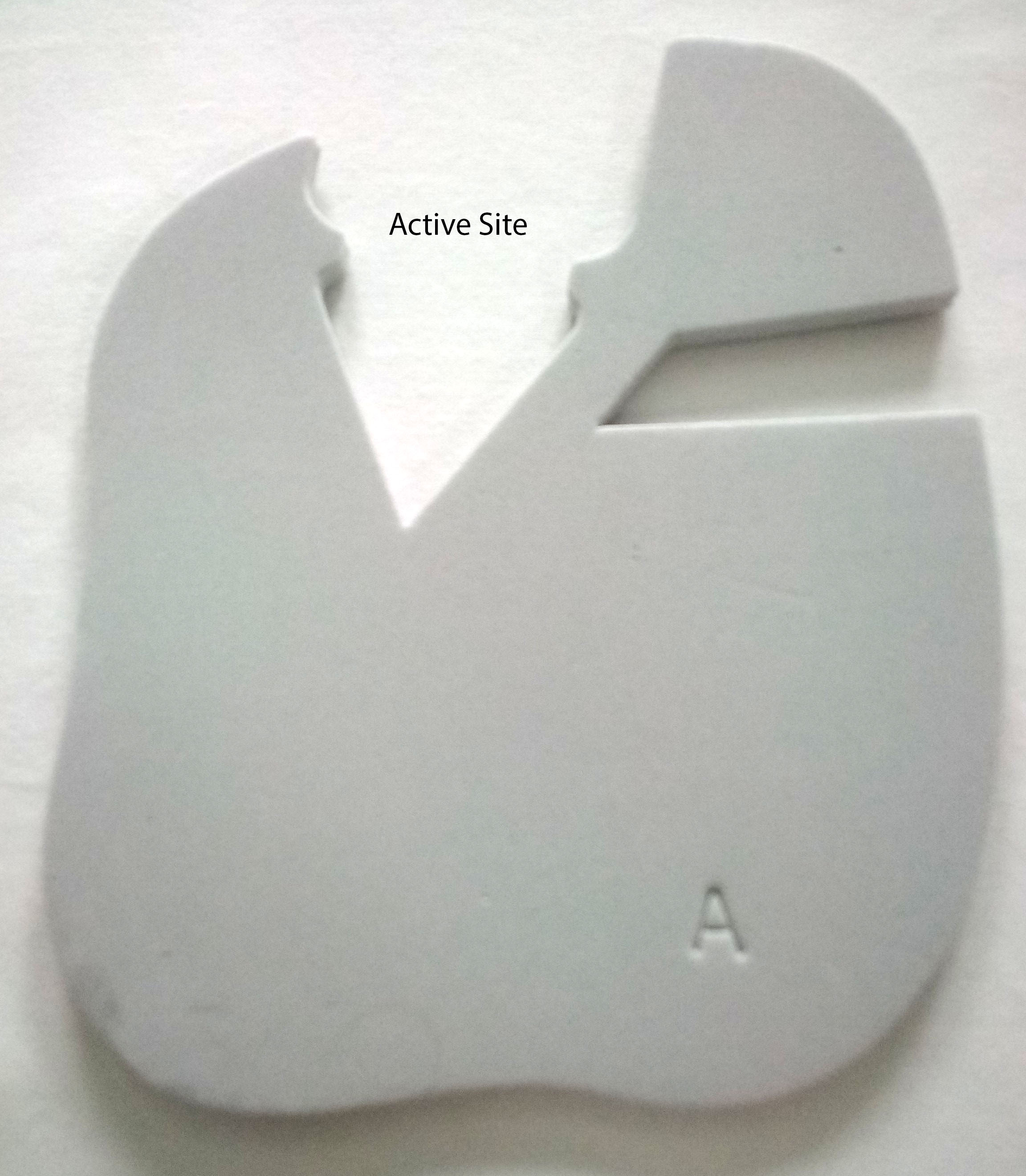
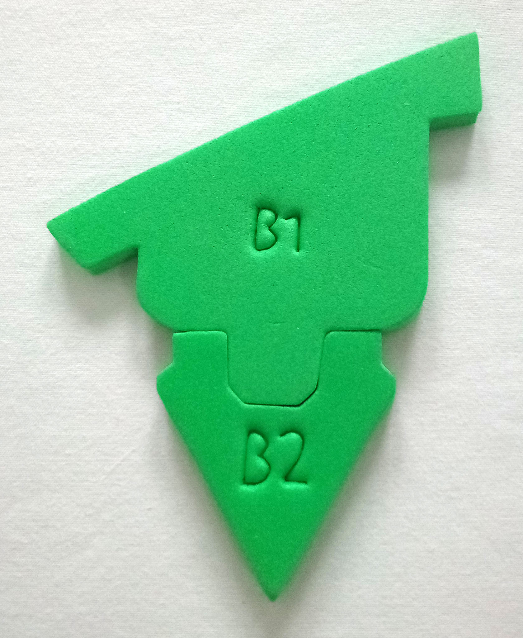
In this model, the gray foam piece represents the enzyme and the two green pieces are the substrate.
1. Are the two substrates pieces bound together before the reaction or not?
Model a catabolic reaction by placing the green piece into the active site of the enzyme. Break the two green pieces apart and place them outside of the enzyme.
2. Did the enzyme change from the beginning to the end of the reaction?
3. Are the two substrates pieces bound together before the reaction or not?
4. Describe how the products are the same/different from the substrate.
5. Does this model better represent the induced fit or lock and key model for enzyme-substrate interactions? Why?
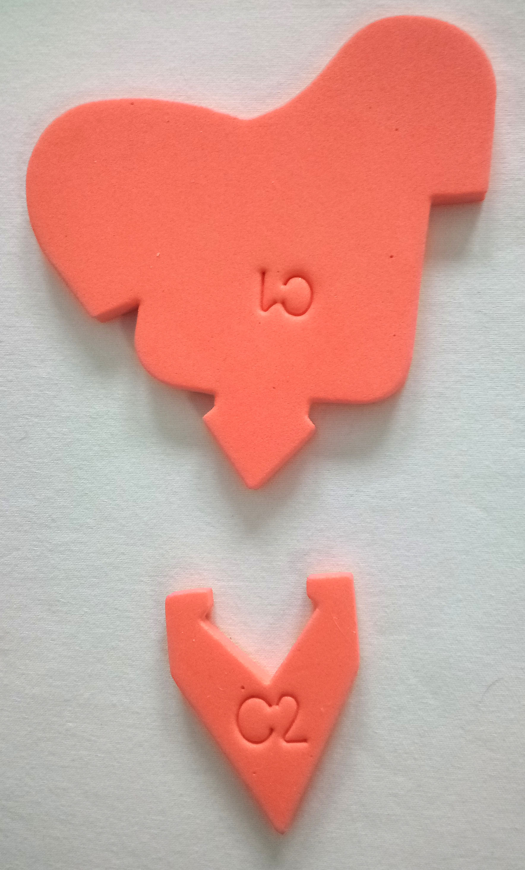
Anabolism is the joining of multiple small molecules to form a larger molecul. The small molecules at the beginning of the reaction are the substrates and the larger molecules after the reaction is the product. You will now use the Enzymes in Action Kit to model anabolism. You will need the following pieces for this activity:
6. Are the two substrates pieces bound together before the reaction or not?
Place the small orange piece into the enzyme active site. Add the larger orange piece to make the final product. Then model the anabolic reaction by putting the two orange pieces together.
7. Describe how the products are the same/different from the substrate.
8. Does this model better represent the induced fit or lock and key model for enzyme-substrate interactions? Why?
9. Write a complete sentence describing anabolism and catabolism.
Anabolism and catabolism are similar words with opposite meaning, so the terms are easily confused. A great pneumonic device is 'Anna had a cat'. If you imagine that Anna is building something (say, a house of cards), and her cat comes along and knocks it down, you have a great way of remembering the difference between these two terms: anabolism is building up, and catabolism is breaking down.
Enzyme specificity is determined by the compatibility between the enzyme and the substrate. The substrate interacts with the active site of the enzyme to bring the substrate into the active site in the correct orientation for catalysis to occur. These attractions are due to noncovalent intermolecular interactions, similar to the ones in protein structure. These include hydrogen bonds (NOT real bonds), salt bridges, and hydrophobic interactions. These interactions are responsible for both a) the substrate and enzyme binding together in the active site and b) the change of the substrate to the product.
To model enzyme specificity, you will need:
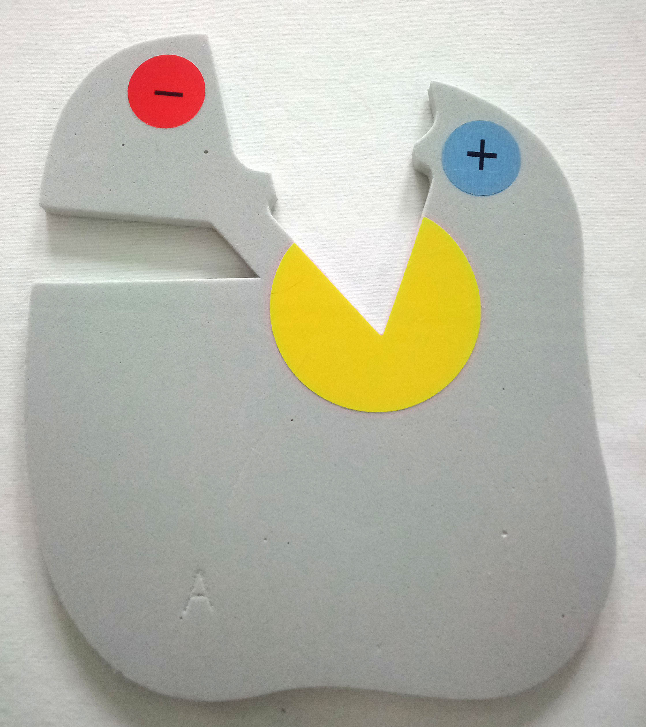
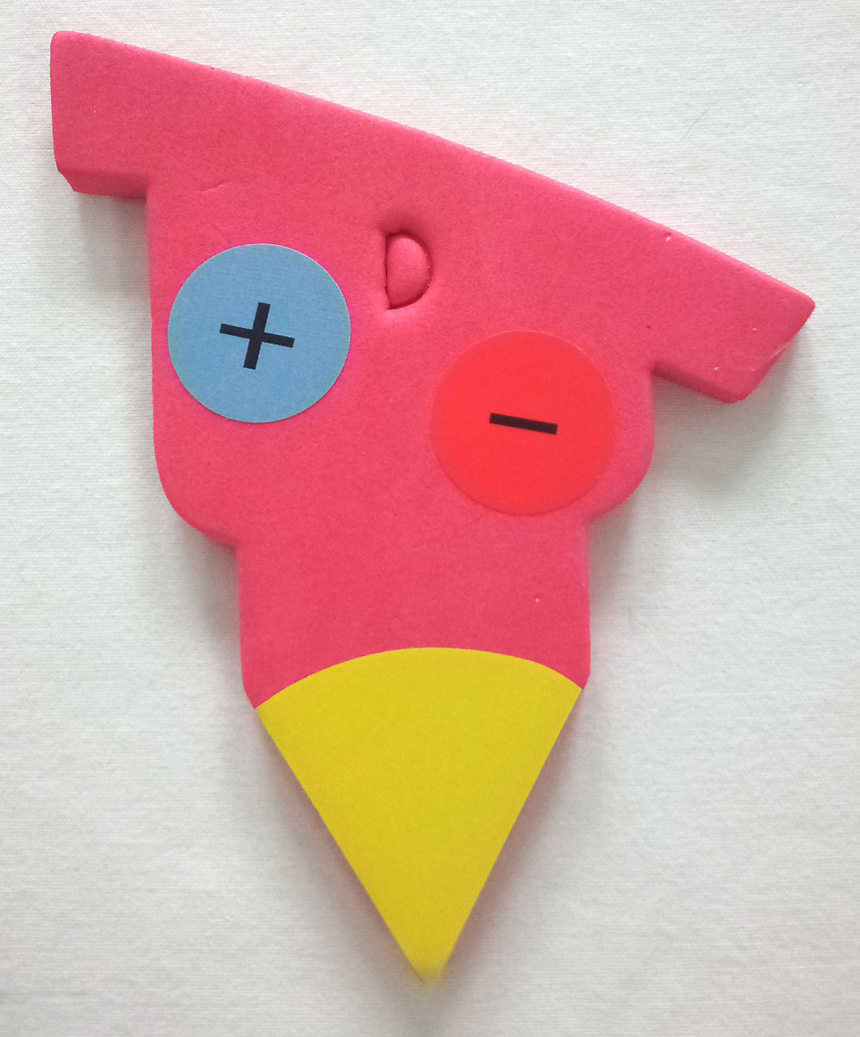
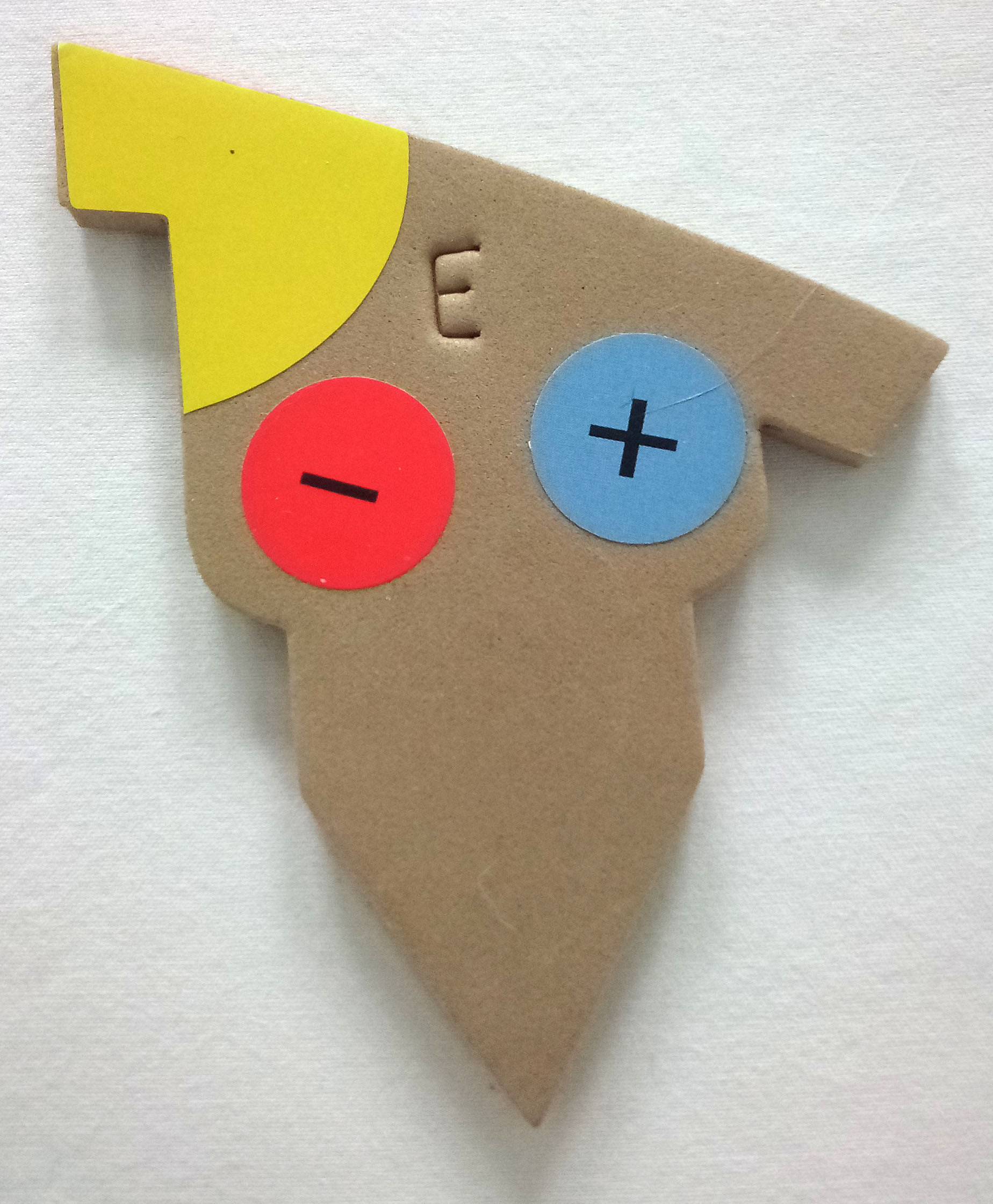
The sticker colors represent chemical characteristics:
10. What are the chemical characteristics of the enzyme's active site?
11. What do the red and tan pieces represent?
12. What are the chemical characteristics of the red and tan pieces?
13. How are the red and tan pieces similar?
14. How are the red and tan pieces different?
Place the red piece in the active site.
15. Will the red piece interact with the enzyme correctly? Why or not?
16. List any interactions that would occur between the enzyme's active site and the red substrate.
17. Will the tan piece interact with the enzyme correctly? Why or not?
18. List any interactions that would occur between the enzyme's active site and the tan substrate.
19. Write a complete sentence describing the two features that make a substrate a good 'fit' in an enzyme active site.
Enzymes are very efficient at catalyzing chemical reactions, but cells often need to regulate enzyme activity so that there is a correct balance of all molecules in the cell. Cells frequently use reversible inhibitors to regulate enzyme activity. These inhibitors can interact with the enzyme to turn it off, but they do not permanently shut off the enzyme.
A simple illustration of this principle is road construction in which there is two-way traffic but only a single lane. Construction workers stand at both ends with a 'stop' and 'proceed with caution' sign that can be flipped. The 'stop' sign is like the enzyme with an inhibitor – cars can't move to the other side. But once enough cars (substrates) build up, and there aren't many cars on the other side (products) the sign is flipped to 'proceed with caution' (inhibitor is removed) and cars can move to the other side (products). The process is reversible because the sign can be flipped 'on' or 'off'.
There is also irreversible inhibition, in which the enzyme is NEVER able to function after interacting with inhibitor. This is analogous to a bridge being washed out in a flood. There is NO traffic until the bridge is rebuilt
In this activity, we will model two forms of reversible enzyme inhibition: competitive inhibition and noncompetitive inhibition.
Competitive inhibition of an enzyme is due to a regulatory molecule that is a similar size and shape of the substrates and does NOT form a product. You will need the following pieces to model competitive inhibition:
Flip the pieces so that the labels are facing the table (and stickers are not visible). For this activity, we will only consider shape of molecules, although chemical characteristics still are important in the binding of substrate and/or inhibitor to the enzyme.
Competitive inhibition occurs when the substrate and the inhibitor are a similar shape, charge, and orientation and will compete for the active site. Place the purple piece in the active site.
20. Can the substrate enter the active site when the competitive inhibitor is present? Why or not?
21.How will the substrate interact with the enzyme's active site?
22. Can the substrate get into the active site once the inhibitor leaves?
Noncompetitive inhibition can also be called allosteric inhibition (from your book). Allosteric enzymes can be either turned on or off by regulatory molecules. Not all allosteric enzymes have both a negative allosteric effector molecule and a positive effector allosteric molecule. Noncompetitive inhibitors DO NOT go into the active site, but into another site on the enzyme. When they interact with the enzyme, they change the shape of the active site. To model noncompetitive inhibition, you will need:
Noncompetitive inhibition occurs when the inhibitor enters the enzyme – not in the active site- and changes the shape of the active site so that the substrate can't enter the active site. To model noncompetitive inhibition, place the blue piece in the other open area (NOT the active site).
23. Can the substrate enter the active site when the noncompetitive inhibitor is present? Why or not?
24. Can the substrate get into the active site once the inhibitor leaves?
25. What would be a purpose for stopping an enzyme from doing its function?