
Serotonin Transporter
This Jmol Exploration was created using the Jmol Exploration Webpage Creator from the MSOE Center for BioMolecular Modeling.
Neurotransmitter transporters are plasma membrane proteins that take up extracellular transmitters and thus stop the transmitters' action at the extracellular receptor sites. Transporters are responsible for the reuptake of neurotransmitters that across the plasma membrane of neurons (Rudnick, 2006). Some of the small neurotransmitters include: dopamine (DA), norepinephrine, and serotonin, which can be transported by proteins and are part of the neurotransmitter sodium symporter (NSS) family. These protein families also contain members from bacterial cells, which can function as amino acid transporters. One of them is the Leucine transporter (LeuT) from Aquifex aeolicus, which shares about 20-25% identity in the primary sequence with neurotransmitter transporters. The crystal structure of the LeuT and its mechanism are good models to study the NSS proteins (Zhou, et al., 2009).
Serotonin transporter (SERT) is a member of the NSS family that transports serotonin (5-hydroxytryptamine, 5-HT) into nerve cells. Once a nerve impulse arrives at an axon, a piece of the nerve cell that sends signals to other cells, neurotransmitters are released to the synapse so they can be received by a specific receptor on the end of a target nerve. After the neurotransmitter is received, it can either excite that nerve to fire its own signal or prevent it from firing its signal (Mandal, 2014). This can also happen with Na + and Cl- and in same reaction also transports a K+ ion out of the cell. SERT can be inhibited by a variety of compounds that are known to treat clinical depression, include sertaline (Zoloft), fluoxetine (Prozac), and paroxetine (Paxil). These compounds are known as selective serotonin reuptake inhibitors (SSRIs) and have become useful antidepressants (Rudnick, 2006).
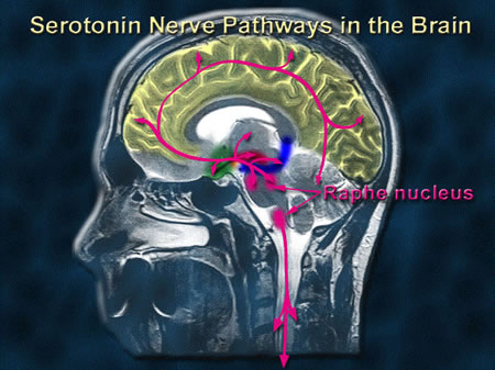
Serotonin is a neurotransmitter located in the brain (fig. 1), but the majority of serotonin is found in the digestive tract. One main function is to help people feel calm, aide in sleep, and maintain a healthy appetite. Those who suffer from low serotonin levels generally suffer from insomnia, depression, or both (Dagnelli, 2013).
There are 515 residues for this protein. It is 72% helical. There are 28 total a-helices (Violet) with 373 residues. There is 1% ?-sheet (yellow) with 4 total strands of sheet and 6 residues.
Beta/AlphaThere are 2 non-protein ligands shown in this structure. They are Sodium (cyan) and Fluoxetine (orange). The latter, fluoxetine (Prozac) interacts with the serotonin transporter in the active site and acts as a competitive inhibitor. Serotonin uptake plays crucial roles in SIDS, Alzheimer's disease, those who suffer from PTSD, OCD, and depression. Prozac works and is affective at blocking the uptake of serotonin in the brain. Blocking the uptake of serotonin in the brain helps brain cells to transmit messages to each other. This allows those suffering from depression or OCD to have a more stable mood (Nordqvist, 2014).
LigandsThis molecule is a transmembrane protein. Due to the nature of transmembrane proteins, resting in a hydrophobic phospholipid bilayer, it is expected that the exterior would be more hydrophobic (yellow) due to complementary hydrophobic residues on the exterior of the protein. The residues shown in red are on the interior of this protein molecule to protect the ligands in the center, which is more hydrophilic. This area is also where serotonin passes.
Hydrophobic/Hydrophilic
Like many other transporters, SERT functions by exposing a substrate-binding site of the cytoplasmic and extracellular of the plasma membrane. SERT then undergoes a conformation change of the binding sites in the extracellular matrix and exposing it to the cytoplasm (Rudnick, 2006). The transporter then returns to its original conformation only after it binds to the K+ ion, which is released to the extracellular medium.
Within leucine (lime green), a drug molecule is able to bind to its extracellular entrance and cause two chlorine atoms on the phenyl ring (fig. 2) to be inserted into a pocket that is formed by Leu25, Gly26, Arg30, Try108, Ile111, and Phe 253. The four halogen-binding residues interact with the opposite side of the polypeptide chain with leucine representing the drug-binding site (Zhou, et al., 2009).

The R-fluoxetine (Prozac) binds to the same extracellular entrance in the LeuT as sertraline (Zoloft). In this case, three fluorines of the methylphenoxy ring is inserted to the same halogen-binding pocket as the two sertraline chlorines that come into contact with Leu25 (dark green), Gly26 (fuchsia), Leu29 (gold), Arg30 (lime green), and Tyr108 (purple). There is also S-fluoxetine, which is the same mirror image as R-fluoxetine (Fig. 3). Due to its opposite chirality, the S-fluoxetine molecule is reversed in the binding pocket (Zhou, et al., 2009).


All of these three SSRIs bind to LeuT in the same position and manner. All have their halogen atoms that are inserted into the same hydrogen binding pockets in LeuT and interact with the same residues. The halogen atoms are important determinates for SSRI specificity, which bind to the same position in LeuT. Mutations at these halogen-binding pockets in SERT will reduce the transporter's affinity for SSRI's. The specificity of the SERT for SSRIs is largely dependent on the interactions that occur with the drug halogens with protein HBP (Zhou, et al., 2009).
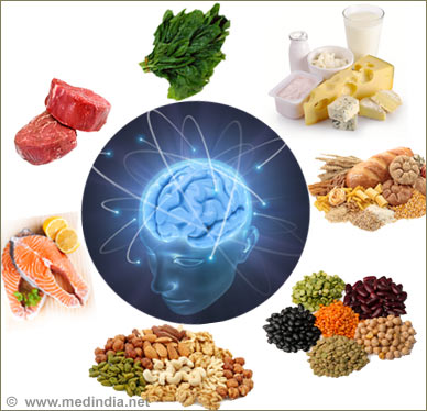
Serotonin rich foods include proteins and oils, fruits and vegetables, and grains. The human body makes serotonin, which is called 5-hydroxytryptophan (5-HTP) from the amino acid tryptophan. We can acquire tryptophan from turkey, eggs, salmon, and sardines.
Serotonin can also exist naturally in kiwi fruit, bananas, plantains, pineapples, and tomatoes. When high amounts of tryptophan in your diet is found to be connected to higher amounts of serotonin levels in the brain. This can only be obtained through your diet as supplements containing high amounts of serotonin does not have the same chemical structure as the serotonin your body makes, therefore, it is unable to be absorbed by the body (Mernitz, 2014).
Dagnelli, C. (2013). Serotonin-rich foods. Retrieved from http://www.livestrong.com/article/261416-serotonin-rich-foods/
Mandal, A. (2014). What is serotonin?. Retrieved from http://www.news-medical.net/health/What-is-Serotonin.aspx
Rudnick, G. (2006). Structure/function relationship in serotonin transporter: new insights from the structure of a bacterial transporter. Yale University School of Medicine, 175, 59-73.
Zhou, Z., Zhen, J., Karpowich, N., Law, C., Reith, M., & Wang, D. (2009). Antidepressant specificity of serotonin transporter suggested by three leut-ssri structures. Nat struct Mol Biol, 16(6), 652-657. doi: 10.1038/nsmb.1602